All hospitals
PrivateCancer Research Hospital

GoBroad Healthcare Group

高博医疗集团

Beijing · Shanghai

Overview

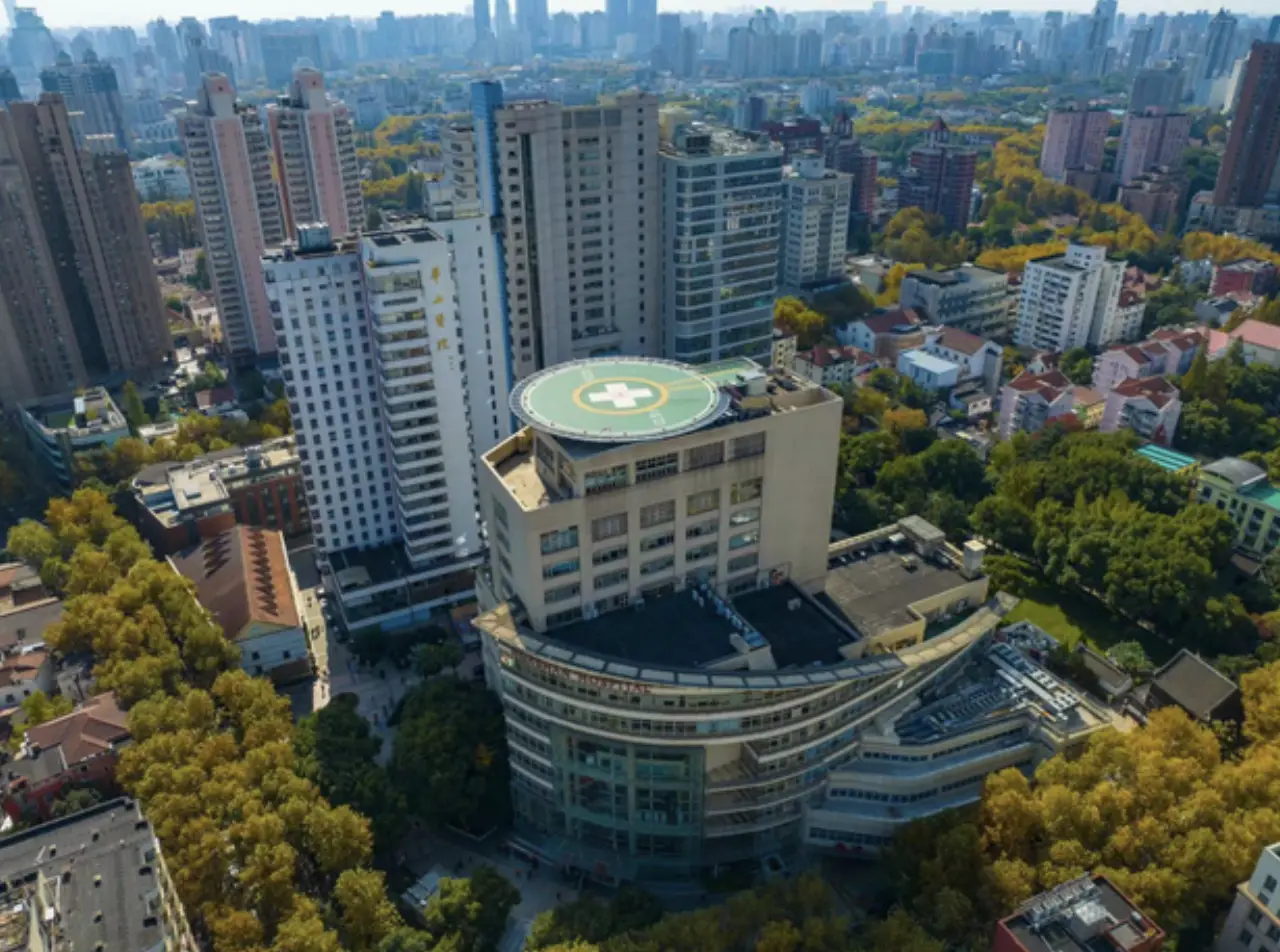
GoBroad Healthcare Group is a research-oriented private hospital group specializing in hematology and oncology, with hospitals in Beijing, Shanghai and Guangzhou and nearly 1,500 open beds. It is one of China's most experienced CAR-T cell therapy centers, having treated more than 5,000 CAR-T patients, including over 1,000 from overseas. The group runs Phase I clinical trial wards, a central laboratory and a dedicated international patient service center.

Why this hospital

Patients' Needs Come First

Advanced equipment

  • Proton Therapy

    Proton therapy, also known as proton beam therapy (PRT), is an advanced radiation therapy technology that uses high-energy proton beams to kill tumor cells. As a cutting-edge tumor treatment method recognized by the international medical community, it relies on the unique "Bragg Peak" physical characteristic of proton beams to achieve precise tumor targeting, with significant advantages in protecting normal tissues and reducing side effects. The specific uses and advantages are as follows: I. Core Uses The indications of proton therapy cover almost all solid tumors suitable for radiation therapy, and it is particularly outstanding in treating tumors in complex locations and special groups of patients. It can be specifically divided into the following two categories: (I) Main Applicable Tumor Types - Central nervous system tumors: Glioma, meningioma, pituitary tumor, arteriovenous malformation, etc. It can significantly reduce the risk of secondary brain tumors after treatment and protect cognitive functions such as memory by reducing the impact on the hippocampus. - Head and neck tumors: Chordoma, nasopharyngeal carcinoma, paranasal sinus/cavity cancer, oropharyngeal cancer, acoustic neuroma, ocular lesions, uveal melanoma, etc. It can help oropharyngeal cancer patients recover normal functions faster and reduce the risk of needing a feeding tube. - Thoracic tumors: Lung cancer, esophageal cancer, breast cancer, thymoma, mediastinal tumor, etc. It can significantly reduce the incidence of radiation esophagitis after treatment. - Abdominal and pelvic tumors: Liver cancer, gastric cancer, pancreatic cancer, prostate cancer, rectal cancer, cervical cancer, bladder cancer, as well as spinal and retroperitoneal sarcomas. - Other tumors: Lymphoma, secondary malignant tumors, malignant skin tumors, sarcomas, as well as bone metastases and lung metastases of various malignant tumors. It can reduce the risk of secondary malignant tumors and wound complications of sarcomas. - Pediatric tumors: Glioma, medulloblastoma, ependymoma, pituitary tumor, ophthalmic tumors, spinal tumors, neuroblastoma, Hodgkin's lymphoma, etc. It can minimize the impact on the developing healthy tissues and organs of children. (II) Key Applicable Scenarios - Tumors in complex locations: Especially suitable for tumors located near important organs (such as eyes, skull base, brainstem) or with irregular shapes, which can maximize the protection of surrounding normal tissues and avoid irreversible damage to important organs. - Re-radiotherapy for recurrent tumors: It can avoid the high-dose damage area of previous radiotherapy, provide a safe treatment path for patients who cannot be treated again with traditional radiotherapy, and reduce the risk of secondary tumors. - Pediatric tumor treatment: As children's bodies are still developing and their organs are closely arranged, proton therapy can significantly reduce radiation to healthy tissues and organs, protect the growth and development of children, and reduce the risk of IQ decline and secondary malignant tumors. - Elderly or weak patients: The treatment process is non-invasive and painless, with low requirements on the patient's physical condition. It is suitable for elderly patients with poor cardiopulmonary function or patients who cannot tolerate or refuse surgery. - Localized tumors: It is particularly effective for localized tumors that have not spread to other parts of the body, and can achieve precise "targeted blasting" of tumor lesions to improve the local control rate of tumors. II. Core Advantages Compared with traditional photon radiotherapy (such as X-ray, gamma-ray) and other advanced radiotherapy technologies, proton therapy takes "precise positioning, minimal damage, and high safety" as its core advantages, which are specifically reflected in the following aspects: (I) Unique Bragg Peak Effect, Precise Dose Distribution The most significant feature of proton therapy is the "Bragg Peak" energy deposition characteristic of proton beams. Before reaching the tumor lesion, the proton beam releases less than 25% of its energy; when it reaches the tumor, it immediately releases the peak energy (Bragg Peak); once it passes through the tumor, the remaining radiation is close to zero. This "targeted sniper" mode can concentrate the radiation dose on the tumor area, realizing precise "blasting" of tumor cells while minimizing the radiation dose to the skin and normal tissues in front of and behind the tumor. (II) Minimal Damage to Normal Tissues, Few Side Effects Compared with traditional radiotherapy and even intensity-modulated radiation therapy (IMRT), proton therapy can significantly reduce the radiation dose received by surrounding normal tissues. For example, in the treatment of head and neck tumors, the radiation dose received by the parotid gland in proton therapy is only 20% of the tumor dose, which is much lower than that of IMRT (33%) and traditional radiotherapy (86%). This can greatly reduce radiation damage, reduce side effects such as dry mouth, nausea, skin reactions, and organ function damage, and significantly improve the patient's quality of life during and after treatment. (III) High Therapeutic Efficacy, Low Recurrence Risk By adjusting the energy of proton beams, the radiation dose in the tumor target area can be maximized (the maximum energy of therapeutic proton beams can reach 250MeV), which can effectively cause double-strand breaks of tumor cell DNA, making it impossible for tumor cells to repair and achieve thorough killing of tumor cells. At the same time, it can reduce the risk of secondary malignant tumors caused by radiation, and expand the treatment methods and means for some advanced tumors, improving the treatment effect and local control rate of tumors. (IV) Good Tolerability, Wide Applicable Population The treatment process of proton therapy is non-invasive and painless, and most patients do not need to be hospitalized (some patients can receive outpatient treatment). It has low requirements on the patient's physical condition, and is suitable for elderly patients, weak patients, children and other special groups that are not suitable for traditional radiotherapy or surgery. Especially for children with tumors, it can protect the normal growth and development of their organs to the greatest extent, reducing the long-term impact of treatment on their physical and mental health. (V) Mature Technology, Flexible Treatment Plans Proton therapy technology has been quite mature in clinical application. It can be divided into proton conformal and intensity-modulated radiotherapy, proton stereotactic radiation therapy, proton scanning irradiation, etc. According to the patient's tumor type, stage, location and physical condition, personalized treatment plans can be customized, and the energy, irradiation angle and treatment cycle of proton beams can be flexibly adjusted to achieve the best treatment effect. At present, domestic proton therapy systems have been put into clinical use, breaking the monopoly of foreign technology and making this advanced treatment technology more accessible. In summary, with its unique Bragg Peak effect, precise dose control and minimal damage to normal tissues, proton therapy, known as the "jewel in the crown of radiotherapy", has become an important advanced treatment option for tumor patients. It not only improves the therapeutic effect of tumors, but also maximizes the protection of normal tissues and organs, taking into account both therapeutic effect and the patient's quality of life, and has broad clinical application prospects.


  • Tomoknife

    Tomoknife, full name Helical Tomotherapy System, is a precise radiotherapy device that integrates spiral CT imaging and intensity-modulated radiation therapy (IMRT) technology. Its core purpose is the radiation treatment of various solid tumors. With its unique technical characteristics, it has significant advantages in tumor treatment. The specific uses and advantages are as follows: I. Core Uses The indications of Tomoknife cover almost all solid tumors suitable for radiation therapy, and it is particularly good at handling complex and refractory tumor scenarios, which can be specifically divided into the following two categories: (I) Main Applicable Tumor Types - Intracranial tumors: Glioma, brain metastases, meningioma, etc. It can accurately avoid important neural structures such as the brainstem and optic nerve, reducing the risk of nerve damage. - Head and neck tumors: Nasopharyngeal carcinoma, laryngeal cancer, tongue cancer, maxillary sinus tumor, etc. It is suitable for re-radiotherapy of recurrent nasopharyngeal carcinoma, reducing complications such as dry mouth and hearing loss. - Thoracic tumors: Lung cancer, esophageal cancer, breast cancer, mediastinal tumor, etc. It can accurately capture the tiny displacement of tumors with breathing to achieve precise irradiation. - Abdominal and pelvic tumors: Liver cancer, pancreatic cancer, prostate cancer, cervical cancer, rectal cancer, etc. It can ensure the therapeutic dose while protecting sensitive organs such as the liver, gastrointestinal tract and bladder. - Other tumors: Spinal tumors, soft tissue tumors, as well as multiple oligometastatic lesions such as bone metastases, lung metastases and lymph node metastases of various malignant tumors. (II) Key Applicable Scenarios - Re-radiotherapy for recurrent tumors: It avoids the high-dose damage area of previous radiotherapy, providing a safe treatment path for patients who cannot be treated again with traditional radiotherapy. - Synchronous treatment of multiple lesions: A single treatment can cover multiple oligometastatic lesions all over the body, without the need for fractional and multi-site treatment, reducing the physical consumption of patients. - Treatment of tumors in complex locations: For tumors close to important organs or with irregular shapes, it maximally protects the surrounding normal tissues and reduces side effects. - Large-scale tumor treatment: It can complete large-scale irradiation such as whole central nervous system and whole bone marrow at one time, with a maximum treatment range of 160cm, far exceeding that of traditional radiotherapy equipment. II. Core Advantages Compared with traditional radiotherapy equipment, Tomoknife takes "precision, safety and efficiency" as its core advantages, which are specifically reflected in the following aspects: (I) Precise Positioning with Minimal Error The equipment is equipped with a built-in MVCT (Megavoltage CT), realizing the integration of "scanning positioning + radiotherapy treatment". Before and during treatment, high-definition images can be obtained in real time, which automatically corrects the patient's positioning error and tumor position deviation (such as displacement caused by breathing), ensuring that the irradiation target area is completely consistent with the tumor. The error can be controlled within 1mm, fundamentally avoiding the mis-irradiation of normal tissues by rays. (II) Precise Dose Regulation and Low Side Effects Equipped with a multi-leaf collimator and intensity-modulated radiation therapy technology, it can accurately adjust the intensity and shape of rays according to the shape and size of the tumor, realizing a gradient distribution of "high dose in the tumor target area and low dose in the surrounding tissues". The local tumor dose can reach 80Gy, which can effectively eliminate tumor cells; at the same time, the radiation dose received by the surrounding normal tissues is 40% lower than that of traditional radiotherapy, greatly reducing radiation damage and side effects such as dry mouth, nausea and organ function damage. (III) Wide Treatment Range and Strong Adaptability The treatment range can cover 160cm, which can not only treat small-volume and precisely positioned tumors, but also complete large-scale irradiation such as whole central nervous system and whole bone marrow; at the same time, it is suitable for a variety of complex tumor scenarios. Whether it is recurrent tumors, multiple metastatic lesions, or tumors with special locations and irregular shapes, effective treatment can be achieved, and the applicable population is wider. (IV) Efficient Treatment and Good Patient Tolerability A single treatment can simultaneously treat multiple lesions all over the body, without the need for repeated fractional and multi-site treatment, shortening the overall treatment cycle; the treatment process is non-invasive and painless, no hospitalization is required (some patients can receive outpatient treatment), the requirements for the patient's physical condition are low, and tumor patients in advanced stages with weak physical condition can also tolerate it well. (V) High Flexibility and Personalized Treatment It can customize personalized treatment plans according to the patient's tumor type, stage and physical condition, and flexibly adjust the irradiation dose, irradiation angle and treatment cycle; at the same time, it supports dynamic tracking of tumor changes and real-time adjustment of treatment parameters, improving the treatment effect and reducing the risk of recurrence. In summary, with its precise positioning, flexible dose regulation and wide adaptability, Tomoknife not only expands the scope of tumor radiotherapy, but also solves the pain points of insufficient precision and obvious side effects of traditional radiotherapy. It has become an important treatment option for patients with complex and advanced tumors, taking into account both treatment effect and patients' quality of life.

  • MRgFUS

    MRgFUS, short for Magnetic Resonance-guided Focused Ultrasound, is an advanced non-invasive therapeutic technology that combines magnetic resonance imaging (MRI) and high-intensity focused ultrasound (HIFU). It uses MRI for real-time positioning, temperature monitoring and navigation, and focuses multiple ultrasound beams on the target area to generate high temperature through thermal effect, thereby ablating abnormal tissues (such as tumors) without incisions or ionizing radiation. As a "green and non-invasive" therapeutic method recognized internationally, it has unique advantages in treating tumors and neurological diseases, with broad clinical application prospects. The specific uses and advantages are as follows: I. Core Uses MRgFUS is mainly applicable to the non-invasive treatment of various benign and malignant solid tumors, neurological diseases and other lesions. With its characteristics of non-invasiveness and high precision, it is widely used in clinical practice, which can be specifically divided into the following two categories: (I) Main Applicable Lesion Types - Neurological diseases: Essential tremor, Parkinson's disease (especially tremor-dominant type), obsessive-compulsive disorder, neuralgia and depression. It is an effective alternative for patients with drug-refractory essential tremor or Parkinson's disease who are not suitable for deep brain stimulation[6][10]. In July 2025, the US FDA further approved it for staged bilateral globus pallidothalamic tract treatment in advanced Parkinson's disease patients. - Gynecological tumors: Uterine fibroids and adenomyosis. It is suitable for intramural, subserosal or submucosal fibroids (FIGO type 2~6) with a maximum diameter of ≤10cm (single or total of multiple fibroids), and can effectively ablate fibroid tissue without damaging normal uterine tissue[5]. - Bone tumors and metastases: Metastatic bone tumors (such as iliac metastasis of alveolar soft tissue sarcoma, lung cancer bone metastasis), which is particularly meaningful for patients with metastatic bone tumors who are insensitive to radiotherapy and chemotherapy or have failed treatment. It can effectively relieve pain and ablate local tumor tissue. - Other tumors: Solid tumors such as liver cancer and breast cancer. It can be used for local ablation of localized tumors, and can also be combined with other therapies to improve the treatment effect. In addition, it can be used in the research of blood-brain barrier disruption to help drugs penetrate the blood-brain barrier and treat central nervous system diseases. (II) Key Applicable Scenarios - Drug-refractory diseases: For neurological diseases such as essential tremor and Parkinson's disease that are ineffective or poorly responsive to drug treatment, MRgFUS can achieve precise ablation of target tissues, significantly improving symptoms with an immediate improvement rate of up to 100% in some cases[10]. - Non-invasive treatment for patients who cannot tolerate surgery: Suitable for elderly patients, patients with poor cardiopulmonary function, or patients who refuse invasive surgery. The whole treatment process is non-invasive, without general anesthesia, and the patient remains awake and can communicate with doctors in real time[10]. - Palliative treatment for advanced tumors: For advanced tumors with distant metastasis (such as metastatic bone tumors), it can effectively relieve tumor-related pain, improve the patient's quality of life, and avoid the side effects of traditional palliative treatment[9]. - Preservation of organ function: When treating uterine fibroids and other gynecological lesions, it can ablate the lesion tissue while preserving the normal structure and function of the uterus, meeting the fertility needs of patients to the greatest extent. - Precise treatment of deep lesions: It can penetrate the skull and other tissues to accurately ablate deep lesions in the brain, with a target accuracy of 0.2~0.7mm, avoiding damage to surrounding normal tissues and important organs. II. Core Advantages Compared with traditional invasive surgery, radiotherapy (such as LINAC, Tomotherapy) and other non-invasive therapies, MRgFUS has the core advantages of non-invasiveness, high precision, safety and quick recovery, which are specifically reflected in the following aspects: (I) Non-invasive and Trauma-free, No Ionizing Radiation MRgFUS does not require incisions, injections or hardware implantation, and the ultrasound beams can penetrate the skin and normal tissues to reach the target area, avoiding surgical trauma, bleeding and infection risks (almost zero infection and bleeding risks). At the same time, it does not use ionizing radiation, which is a "green and safe" therapeutic method that can avoid radiation damage to normal tissues and reduce the risk of secondary tumors. (II) Real-time MRI Guidance, High Precision and Safety The integration of MRI technology enables real-time positioning, navigation and temperature monitoring during treatment. MRI can accurately display the location, shape and size of the lesion, and monitor the temperature of the target area in real time (controlling the ablation temperature at 60~100℃), so as to adjust the focus position and energy in time, ensuring that the ultrasound beams are accurately focused on the lesion area while minimizing damage to surrounding normal tissues[5][9]. (III) Quick Recovery and Less Impact on Quality of Life The treatment process is short (usually 1~1.5 hours), and patients can be discharged the next day after treatment, with a hospital stay of less than 24 hours in most cases. There is no obvious discomfort during treatment, and there is no need for long-term recovery after treatment, which greatly reduces the physical and mental burden of patients and has little impact on their daily life and work. (IV) Flexible Treatment and Strong Individualization According to the patient's lesion type, location, size and physical condition, the focus position, energy and ablation range of ultrasound beams can be flexibly adjusted to formulate personalized treatment plans. It can not only treat single lesions, but also target multiple lesions, and can also be combined with other therapeutic methods to improve the overall treatment effect. (V) Mature Technology and Wide Clinical Recognition MRgFUS has obtained the authoritative certification of NMPA in China, FDA in the United States and CE in the European Union, and has been clinically applied in more than 200 medical centers around the world, with more than 25,000 patients treated cumulatively. Its clinical safety and effectiveness have been fully verified, and it has become an important non-invasive treatment option in the field of tumors and neurological diseases. In summary, as an advanced non-invasive therapeutic technology, MRgFUS integrates the advantages of MRI's precise positioning and focused ultrasound's non-invasive ablation. It has the characteristics of non-invasiveness, high precision, safety and quick recovery, providing a new treatment path for patients with drug-refractory neurological diseases, tumors and other lesions. It not only improves the treatment effect, but also maximizes the protection of normal tissues and organ functions, which has important clinical significance and broad application prospects.
  • LINAC

    LINAC, short for Linear Accelerator, is a core device in modern radiation therapy that generates high-energy X-rays or electron beams to kill tumor cells. As the most widely used and mature radiation therapy equipment in clinical practice, it has strong adaptability, flexible operation and high cost-effectiveness, playing an irreplaceable role in tumor treatment. The specific uses and advantages are as follows: I. Core Uses LINAC is applicable to the radiation treatment of most solid tumors and part of hematological tumors, and is widely used in clinical oncology due to its flexible treatment modes. It can be specifically divided into the following two categories: (I) Main Applicable Tumor Types - Head and neck tumors: Nasopharyngeal carcinoma, laryngeal cancer, tongue cancer, oropharyngeal cancer, maxillary sinus tumor, acoustic neuroma, etc. It is the first-choice radiotherapy equipment for most head and neck tumors, especially for early and mid-stage lesions. - Thoracic tumors: Lung cancer (non-small cell lung cancer, small cell lung cancer), esophageal cancer, breast cancer, thymoma, mediastinal tumor, etc. It can be used for radical radiotherapy, adjuvant radiotherapy and palliative radiotherapy of thoracic tumors. - Abdominal and pelvic tumors: Liver cancer, gastric cancer, pancreatic cancer, colorectal cancer, prostate cancer, cervical cancer, endometrial cancer, bladder cancer, etc. It is suitable for local treatment of localized tumors and palliative treatment of advanced tumors. - Central nervous system tumors: Brain glioma, brain metastases, meningioma, pituitary tumor, etc. It can be combined with stereotactic radiotherapy technology to improve the treatment precision of intracranial tumors. - Other tumors: Skin cancer, soft tissue sarcoma, bone tumors, lymphoma, as well as bone metastases, lung metastases and lymph node metastases of various malignant tumors. Electron beam irradiation of LINAC is particularly suitable for superficial tumors such as skin cancer. - Pediatric tumors: Common pediatric malignant tumors such as neuroblastoma, Wilms tumor, medulloblastoma, and Hodgkin's lymphoma. It can adjust the radiation dose and field according to the child's physical development to reduce side effects. (II) Key Applicable Scenarios - Radical radiotherapy: For early and mid-stage localized tumors (such as early lung cancer, nasopharyngeal carcinoma, prostate cancer), LINAC can deliver sufficient radiation dose to the tumor target area to achieve radical cure, with a high local control rate. - Adjuvant radiotherapy: After surgery or chemotherapy, it is used to eliminate residual tumor cells, reduce the risk of tumor recurrence and metastasis, and improve the overall treatment effect (such as adjuvant radiotherapy after breast cancer and colorectal cancer surgery). - Palliative radiotherapy: For advanced tumors with distant metastasis, it can relieve tumor-related symptoms (such as pain, obstruction, bleeding), improve the patient's quality of life, and prolong the survival period. - Stereotactic radiotherapy: Equipped with image-guided radiotherapy (IGRT) and intensity-modulated radiation therapy (IMRT) functions, LINAC can be used for stereotactic body radiation therapy (SBRT) and stereotactic radiosurgery (SRS), suitable for small tumors and oligometastatic lesions. - Superficial tumor treatment: The electron beam generated by LINAC has the characteristics of shallow energy deposition, which is especially suitable for the treatment of superficial tumors such as skin cancer and superficial soft tissue sarcoma, avoiding damage to deep normal tissues. II. Core Advantages Compared with other radiation therapy equipment (such as proton therapy, Tomotherapy), LINAC has the advantages of mature technology, wide application range, high cost-effectiveness and flexible operation, which are specifically reflected in the following aspects: (I) Mature Technology and High Clinical Applicability LINAC has a history of more than 60 years of clinical application, and its technology is extremely mature and stable. It has been widely popularized in hospitals at all levels around the world, and the treatment plan and operation process have been standardized. It can meet the treatment needs of most tumor patients, and the clinical safety and effectiveness have been fully verified. (II) Flexible Treatment Modes and Strong Adaptability LINAC can generate two types of radiation beams: X-rays and electron beams, which can be flexibly selected according to the location, depth and size of the tumor. X-rays are suitable for deep tumors, while electron beams are suitable for superficial tumors. At the same time, it can be combined with IMRT, IGRT, SBRT and other advanced technologies to realize personalized treatment for different patients and tumor types. (III) High Cost-Effectiveness and Wide Accessibility Compared with advanced radiotherapy technologies such as proton therapy, the construction and operation costs of LINAC are much lower, and the treatment cost is more affordable for patients. It has been widely popularized in primary, secondary and tertiary hospitals, making radiation therapy accessible to more tumor patients, especially in developing regions and areas with limited medical resources. (IV) Precise Irradiation and Controllable Side Effects Modern LINAC is equipped with high-precision multi-leaf collimators and image-guided systems (such as CBCT), which can accurately locate the tumor, adjust the radiation field and dose according to the shape of the tumor, and minimize the radiation dose to surrounding normal tissues. While ensuring the therapeutic effect, it effectively controls radiation side effects and improves the patient's quality of life during treatment. (V) Simple Operation and Short Treatment Cycle The operation process of LINAC is relatively simple, and the treatment time per session is short (usually 10-20 minutes), which reduces the physical and mental burden of patients. For most patients, the treatment cycle is 4-7 weeks, which is shorter than some advanced radiotherapy technologies, and the compliance of patients is higher. In summary, as the most widely used radiation therapy equipment in clinical practice, LINAC has the characteristics of mature technology, wide application range, high cost-effectiveness and flexible operation. It is the cornerstone of tumor radiation therapy, providing effective treatment options for a large number of tumor patients, and playing an important role in improving the treatment effect of tumors and prolonging the survival period of patients.

  • PET-CT

    PET-CT, short for Positron Emission Tomography-Computed Tomography, is an advanced integrated imaging technology that combines positron emission tomography (PET) and computed tomography (CT). It integrates the functional metabolic information of PET (reflecting the physiological and metabolic state of tissues) with the anatomical structural information of CT (showing the location, shape and size of lesions), realizing "functional + structural" dual-mode imaging. As a core tool in modern clinical oncology, cardiovascular disease and neurological disease diagnosis, it has unique advantages in early detection, accurate positioning and etiological diagnosis. The specific uses and advantages are as follows: I. Core Uses PET-CT is mainly used for the diagnosis, staging, treatment evaluation and follow-up of tumors, as well as the diagnosis and evaluation of cardiovascular and neurological diseases. With its dual-mode imaging advantage, it provides comprehensive and accurate diagnostic information for clinical practice, which can be specifically divided into the following two categories: (I) Main Applicable Disease Types - Tumor-related diseases: It is widely used in the diagnosis and management of various malignant tumors, including lung cancer, breast cancer, lymphoma, colorectal cancer, pancreatic cancer, liver cancer, esophageal cancer, prostate cancer, ovarian cancer, brain tumors, and metastatic tumors of various origins. It can identify benign and malignant lesions, clarify the scope of tumor invasion and distant metastasis, and provide a basis for formulating treatment plans. - Cardiovascular diseases: Myocardial ischemia, myocardial infarction, cardiomyopathy, and evaluation of myocardial viability. It can accurately assess the blood supply and metabolic state of the myocardium, determine the location and extent of myocardial ischemia or infarction, and guide the selection of treatment methods (such as interventional therapy or surgical treatment). - Neurological diseases: Alzheimer's disease (senile dementia), epilepsy, Parkinson's disease, cerebrovascular diseases (such as cerebral infarction), brain tumors and mental disorders. It can detect abnormal changes in brain metabolism in the early stage, assist in distinguishing the type and cause of neurological diseases, and evaluate the severity of lesions and treatment effects. - Other applications: It can also be used for the diagnosis of infectious diseases (such as tuberculosis, fungal infections) and inflammatory diseases, helping to locate the focus of infection or inflammation and clarify the scope of lesions. (II) Key Applicable Scenarios - Early tumor detection and screening: For high-risk groups of tumors (such as long-term smokers, family history of tumors), PET-CT can detect abnormal metabolic lesions that are not yet manifested in anatomical structure, realizing early discovery, early diagnosis and early treatment of tumors, which is particularly important for improving the survival rate of tumor patients. - Tumor staging and treatment plan formulation: It can accurately determine the stage of tumors (primary lesion, regional lymph node metastasis, distant metastasis), clarify whether the tumor has spread, and help clinicians formulate personalized treatment plans (such as surgery, radiotherapy, chemotherapy, targeted therapy). - Evaluation of tumor treatment effect: After surgery, radiotherapy, chemotherapy or targeted therapy, PET-CT can evaluate whether the tumor is completely removed, shrunk, stable or progressed, judge the curative effect in time, and adjust the treatment plan if necessary. - Search for primary tumor of unknown origin: For patients with metastatic tumors but unknown primary lesion, PET-CT can comprehensively scan the whole body to find the primary tumor, providing a basis for etiological diagnosis and treatment. - Evaluation of myocardial viability and neurological function: For patients with myocardial infarction, it can evaluate whether the damaged myocardium has viability, guiding the selection of revascularization therapy; for patients with epilepsy, it can locate the epileptic focus, providing a basis for surgical treatment. II. Core Advantages Compared with single PET, single CT and other imaging technologies (such as MRI), PET-CT has the advantages of comprehensive information, high diagnostic accuracy, early detection and one-stop inspection, which are specifically reflected in the following aspects: (I) Dual-mode Integration, Comprehensive Diagnostic Information PET-CT combines the functional metabolic information of PET and the anatomical structural information of CT. PET can detect abnormal metabolic activities of lesions (such as increased glucose metabolism in tumor cells) in the early stage, while CT can accurately locate the lesions and show their anatomical relationship with surrounding tissues. The combination of the two avoids the limitations of single imaging technology, providing more comprehensive and accurate diagnostic information for clinicians. (II) High Diagnostic Accuracy, Reducing Misdiagnosis and Missed Diagnosis By integrating functional and structural information, PET-CT can effectively distinguish benign and malignant lesions, which is significantly higher than single PET or CT in the diagnosis of early tumors, metastatic tumors and complex lesions. It can reduce the rate of misdiagnosis and missed diagnosis, avoid unnecessary invasive examinations (such as biopsy) and improper treatment, and improve the level of clinical diagnosis. (III) Early Detection of Lesions, Leading to Early Treatment PET-CT can detect abnormal metabolic changes of lesions before obvious anatomical structural changes occur (usually 3-6 months earlier than traditional imaging technologies). This early detection advantage is particularly important for tumors, cardiovascular diseases and neurological diseases, which can help patients get early treatment, improve the treatment effect and reduce the mortality rate. (IV) One-stop Whole-body Inspection, High Efficiency PET-CT can complete whole-body imaging in one inspection, without the need for multiple separate inspections (such as separate PET and CT). It saves inspection time, reduces the burden on patients, and can comprehensively evaluate the whole body situation, especially for the detection of systemic diseases (such as metastatic tumors) and the search for primary lesions of unknown origin. (V) Guiding Treatment and Evaluating Prognosis, Strong Clinical Guidance PET-CT can not only assist in diagnosis and staging, but also guide the formulation of treatment plans, evaluate the treatment effect and predict the prognosis of patients. For tumor patients, it can help clinicians judge whether the treatment is effective, adjust the treatment plan in time, and improve the survival rate and quality of life of patients; for cardiovascular and neurological disease patients, it can provide a basis for treatment selection and evaluate the recovery of disease. In summary, as an advanced integrated imaging technology, PET-CT integrates the advantages of PET and CT, with the characteristics of comprehensive information, high accuracy, early detection and high efficiency. It has become an important diagnostic tool in clinical practice, playing a key role in the diagnosis, treatment and follow-up of tumors, cardiovascular diseases and neurological diseases, and providing strong support for improving the level of clinical medical treatment.

Frequently asked questions

Common questions from international patients

Compiled from the hospital's official website and other public sources. Details may change, so please confirm with us before you travel.

More hospitals in Beijing

Raffles Hospital Beijing
Private
Beijing

Raffles Hospital Beijing

北京莱佛士医院

Raffles Hospital Beijing is part of Raffles Medical Group, a leading integrated private healthcare provider in Asia founded in Singapore in 1976. The group serves more than 2.8 million patients a year and operates medical facilities in 12 cities across Singapore, Greater China, Japan, Vietnam and Cambodia. Its Beijing facility offers international-standard care with multilingual staff.

  • Est. 1996
View details
Eye & ENT Hospital of Fudan University
Grade 3APublic
Shanghai

Eye & ENT Hospital of Fudan University

复旦大学附属眼耳鼻喉科医院

Eye & ENT Hospital of Fudan University (复旦大学附属眼耳鼻喉科医院) was established in 1952 in Shanghai, as a Grade 3A comprehensive hospital, with professional advantages in Cataract and Refractive Surgery Center, Cornea and Ocular Surface Disease Center, Glaucoma Center and other fields.

  • Est. 1952
  • 12 departments
  • 3.3M visits/yr
View details
Huashan Hospital, Fudan University
Grade 3APublic
Shanghai

Huashan Hospital, Fudan University

复旦大学附属华山医院

Huashan Hospital, Fudan University (复旦大学附属华山医院) was established in 1907 in Shanghai, as a Grade 3A comprehensive hospital, with professional advantages in Neuromedicine Center (Neurosurgery/Neurology/Neuro-ICU), Cerebrovascular and Stroke Center, Functional Neurosurgery and Neuromodulation Center and other fields.

  • Est. 1907
  • 10 departments
  • 6M visits/yr
View details